Reproduction is one of the basic characteristics of life phenomena, and is the process of life continuation between parents and offspring. It can be divided into female reproductive basic and male reproductive diseases, including polycystic ovarian syndrome, uterine adhesion, vasectomy, neurogenic erectile dysfunction, etc. However, the reproductive system characteristics and reproductive physiological processes of animals and humans are different, and the symptoms of related diseases on human and animal kidneys are naturally different. Therefore, there are some limitations in the relevant models, such as rabbits have no menstrual cycle.
Pregnancy induced hypertension model
L-NAME induced pregnancy induced hypertension model in rats
Polycystic ovary syndrome model
Establishment of rat model of polycystic ovary syndrome by high fat diet combined with subcutaneous injection of DHEA
Establishment of rat model of polycystic ovary syndrome with high-fat diet and LZ
Thin uterus model
Establishment of thin uterus model by injecting 95% ethanol into uterine horn
Endometriosis model
Establishment of endometriosis model by transferring uterine horn to abdominal wall of mice
Establishment of rat endometriosis analgesia model by wrapping endometrium in sciatic nerve
Uterine adhesion model
Establishment of rabbit uterus adhesion model by curettage combined with alcohol injury
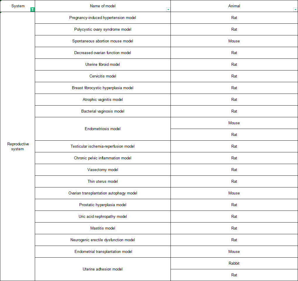
Prostatic hyperplasia model
Establishment of a rat model of benign prostatic hyperplasia by orchiectomy combined with subcutaneous injection of testosterone propionate
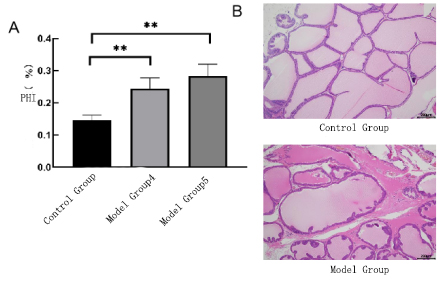
Fig. 2 Prostatic hyperplasia model. A Prostate index, indicating that the prostate index in the model group increased significantly. B HE staining showed that in the model group, the glandular epithelium was obviously hyperplasia, fibrous tissue hyperplasia and inflammatory cell infiltration were visible in the stroma
Mastitis model
Establishment of mastitis model by injecting LPS into breast
(Control Group) (Model Group)
Fig. 3 Mastitis model. The HE of breast tissue showed that in the model group, the acinar space widened, the connective tissue proliferated, the acinar interstitium bled, the acinar epithelial cells necrosis, and a large number of inflammatory cells gathered in the acinar cavity.