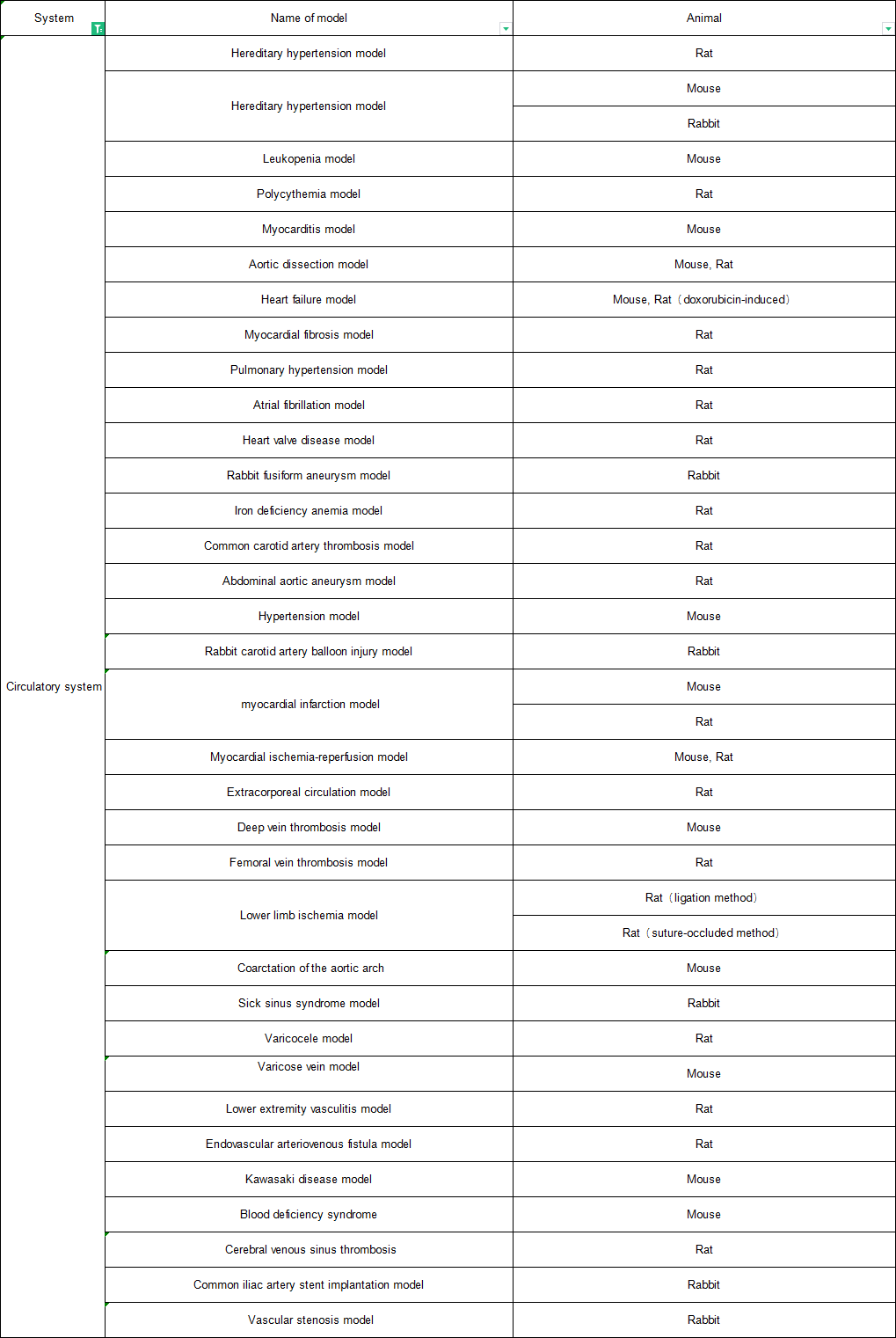
Cardiac and vascular diseases and their complications are complex multifactorial pathological processes, and are the result of the joint action of genes and environmental factors. Animals have inestimable value in the research of cardiovascular system. Many cardiovascular disease models have been successfully replicated in different species, such as myocardial infarction models in rats and rabbits, and atherosclerosis models in rabbits.
Hypertension model
Spontaneously hypertensive (SHR) rats
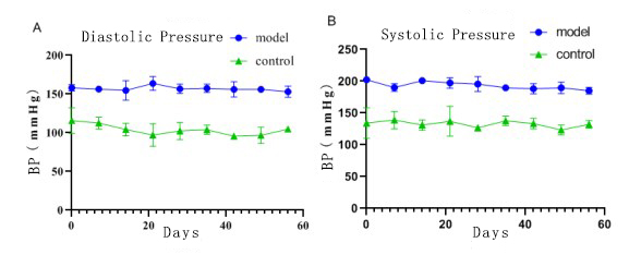
Fig. 1 Hypertension model. The blood pressure results showed that the diastolic and systolic blood pressure in the model group were higher than those in the control group.
Atherosclerosis model
ApoE -/- gene knockout mice
Atherosclerosis model induced by high fat diet in rabbits
Autoimmune myocarditis model
Establishment of mouse autoimmune myocarditis model by injecting myosin emulsion
Aortic dissection model
β- Establishment of mouse aortic dissection model by adding aminopropionitrile into drinking water and intraperitoneal injection of Ang - Ⅱ
(control group) (Model Group)
Figure 2 Aortic dissection model. HE and MASSON staining of aorta showed obvious dissection in model group.
Heart failure model
Adriamycin induced heart failure model in rats
Atrial fibrillation model
Establishment of rat atrial fibrillation model by esophageal electrical stimulation
Abdominal aortic aneurysm model
Establishment of rat abdominal aortic aneurysm model by elastase perfusion
Rabbit carotid artery balloon injury model
Establishment of rabbit model of common carotid artery balloon injury by pressure pump injury
Myocardial infarction model
Establishment of myocardial infarction model in rats/mice by ligation of left anterior descending coronary artery
Myocardial ischemia reperfusion model
Establishment of myocardial infarction model in rats/mice by releasing the left anterior descending coronary artery after ligation
(control group)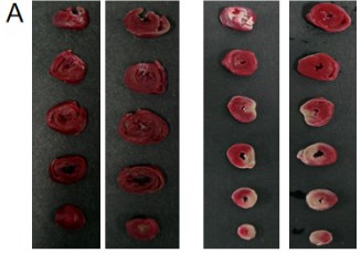 (Model Group)
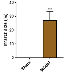
(control group)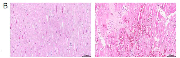 (Model Group)
Fig. 3 Myocardial ischemia reperfusion model. A TTC staining. The infarct area of myocardial tissue in the model group increased significantly. B HE staining of myocardial tissue. The myocardial tissue in the model group showed obvious pathological changes, including erythrocyte exudation and myocardial fiber rupture.
Cardiopulmonary bypass model
Establishment of extracorporeal circulation model by jugular blood flowing back to carotid artery through peristaltic pump
Aortic arch constriction model
Establishment of aortic arch constriction model by ligation of constricted aortic arch
Inferior vena cava stenosis model
Establishment of inferior vena cava stenosis model by suture ligation and vascular clamping
Sick sinus syndrome model
Establishment of sick sinus syndrome model in rabbits by wet compress of sinus node with formaldehyde
Myocardial fibrosis model
Establishment of rat myocardial fibrosis model by subcutaneous injection of isoproterenol hydrochloride
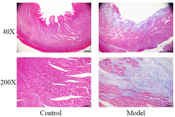
Fig. 4 Myocardial fibrosis model. HE staining pictures of myocardial tissue showed that a large number of muscle fibers were damaged and fibrosis was serious in the model group.
Lower limb vasculitis model
Establishment of vasculitis model of lower limbs in rats by sodium laurate injection
Model of arteriovenous bypass vascular fistula
Establishment of arteriovenous bypass graft fistula model by sodium butyrate injection combined with femoral vein catheterization