The animal model of skeletal system is the premise and basis for the research of bone related diseases, fracture and bone defect repair, as well as orthopedic-related repair materials and internal fixation devices. A large number of new products and new technologies have been widely used in orthopedic clinic, and all of these achievements can not be achieved without the early application of basic research, in which the animal model is an important research tool.
Osteoarthritis model
Cutting the anterior cruciate ligament of the knee joint to establish knee arthritis in rats/mice
Osteoarthritis induced by local injection of 1% papain and L-cysteine solution in rabbits
Fig. 1 Rat osteoarthritis model. Schematic diagram of cutting the anterior cruciate ligament of the knee joint.
Gouty arthritis model
Rat model of gouty arthritis induced by sodium urate crystal injection
(Before molding)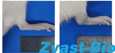 (After molding)
Fig. 2 Rat model of gouty arthritis. The swelling of the ankle joint at the back of the model was obvious.
Ovariectomized osteoporosis model
Establishment of ovariectomized osteoporosis model in rats by bilateral ovariectomy
Fracture model
Establishment of tibial fracture model in rats by intramedullary Kirschner wire fixation after tibial fracture
Radial fracture model
Model of lumbar intervertebral disc degeneration
Stress imbalance caused by cutting the joint connection of thoracic vertebrae induces lumbar intervertebral disc degeneration in rats
Complete spinal cord transection model
Establishment of complete spinal cord transection model in rats
Femoral head necrosis model
Establishment of rat femoral head necrosis model by injecting methylprednisolone
(control group)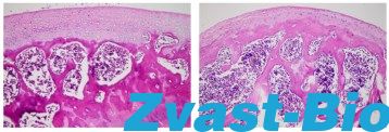 (Model Group)
Fig. 3 Rat model of femoral head necrosis. The bone tissue in the model group was damaged to a certain extent, of which the bone trabecular structure was changed most obviously and arranged in disorder, accompanied by slight fracture of bone trabecula, decreased hematopoietic cells in the bone marrow cavity, obvious thickening of bone trabecula, and cartilage lesions, suspected fibrosis, and a small amount of oil cells were visible under the microscope.
Bone defect model
Model of large segmental bone defect of radius
Model of femoral cavernous bone defect
Mandibular defect model