Language

 视网膜感光细胞,动物造模
 z-bo
z-bo
 2020-07-24
 1548
1548
According to reports, the pig retina contains a large number of mitochondria. Since there are few reports of large mitochondria in mammalian photoreceptors, scanning electron microscopy (SEM) was used to evaluate the morphology of pig, rabbit, and mouse retinas. In order to obtain high-resolution images with a wide field of view, we have adopted a unique scanning electron microscope method. With traditional SEM, no embedding and slicing steps are required, only the surface of the specimen is visible. In contrast, our SEM technique involves mounting single or continuous ultrathin sections on a glass slide, staining with heavy metals and scanning backscattered electrons. By digitally "combining" continuous SEM images, images with a wide field of view can be acquired with high resolution. Figure 1a shows an electron microscope image of the porcine retina. Figure 1b is an electron microscope image of the porcine photoreceptor cell layer. Fig. 1c is an enlarged view of the viewing cone in Fig. 1a, Fig. 1d is an enlarged view of the rod in Fig. 1a, and Fig. 2a is an electron microscope image of the rabbit retina. Figure 2b is an electron microscope image of the rabbit photoreceptor layer. Figure 2c shows an enlarged view of the frustum of Figure 2a. Figure 2d shows an enlarged view of the rod portion of Figure 2a. Figure 3a shows an electron microscope image of the mouse retina. Figure 3b shows an electron microscope image of the mouse photosensitive layer. Figure 3c shows an enlarged view of the viewing cone of Figure 3a. Fig. 3d shows an enlarged view of the rod part of Fig. 3a. OS: external, IS: internal, ONL: external nuclear layer, OPL: external plexiform layer, INL: internal plexiform layer, IPL: internal plexiform layer, GCL: ganglion cell layer. CIS: Cone optical receiver. IS: Rod sensor part.
Experimental design, materials, methods
The retina plays an important role in achieving vision, because it participates in receiving light stimulation and sending signals to the visual cortex. The vertebrate retina is composed of several cell layers, including ganglion cells, bipolar cells, amacrine cells, horizontal cells and photoreceptor cells. In addition, the photoreceptor exhibits functional specificity. Rod-shaped photoreceptors mediate scotopic vision, and cone cells mediate photopic vision. It takes a lot of energy to absorb light and convert it into electrical signals (photoelectric conversion) and to transmit the signals from the photoreceptor to the retinal neurons. To meet this high energy demand, many mitochondria are distributed in the inner region of the photoreceptor. In some species, such as tiger head fish and zebrafish, the inner part of the cone photoreceptor contains heterogeneous mitochondria with a diameter greater than 2 μm, called giant mitochondria. It is generally believed that cone photoreceptors consume more energy than rod photoreceptors, and huge mitochondria are responsible for the high energy production of these cells. However, the physiological significance of these characteristic mitochondria is not fully understood. In our previous study, the porcine retina also contained large mitochondria similar to those observed in zebrafish, which only clustered within the pyramidal photoreceptor cells and clustered with each other. .. Further research on other mammalian species is needed to clarify the biological significance of the existence of giant mitochondria in photoreceptors. We observed the retinas of pigs, rabbits and mice. The original SEM method is used to obtain high-resolution images with a wide field of view. In this experiment, electron microscopy was used to examine the presence of giant mitochondria in the inner part of photoreceptor cells in the rabbit and mouse retina. We also compared the mitochondrial morphology of the inner part of photoreceptor cells in pig, rabbit, and mouse retinas.
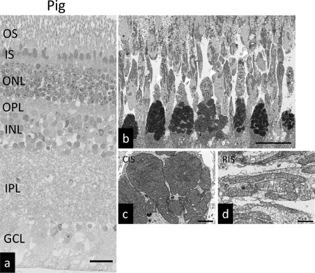
图1.猪视网膜的超微结构。
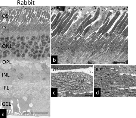
图2.兔子视网膜的超微结构。
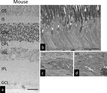
图3.小鼠视网膜的超微结构。
Animal: Pig eyes from slaughterhouse. Eight-week-old New Zealand rabbits were euthanized with isoflurane, and the eyeballs were removed to detach the retina. Eight-week-old C57BL/6 mice were euthanized with isoflurane, the fixative was perfused, the eyeballs were removed, and the retinas were removed.
Use a scanning electron microscope to observe the morphology of the retina. With a conventional scanning electron microscope, no embedding and slicing steps are required, only the surface is observed. The SEM technique used in this experiment involves mounting single or continuous ultrathin sections on a glass slide, staining them with heavy metals, and scanning backscattered electrons. By digitally "combining" continuous SEM images, images with a wide field of view can be acquired with high resolution. The anterior structure of the cornea, lens and iris was removed at 4°C and removed with a fixative (2.5% glutaraldehyde, 2% paraformaldehyde solution, 0.1M sodium phosphate buffer, pH 7.4). I will stay overnight. Rinse the eyes twice with 0.1 M phosphate buffer for 10 minutes each time, and then chop the retina. The trimmed retina was fixed in 1% tetroxide for 2 hours, and then dehydrated in various concentrations of ethanol (50%, 70%, 80%, 90%, 100%) for 15 minutes. After dehydration, the sample was rinsed twice with QY-1 (15 minutes each time), rinsed once with a mixture of QY-1 and Epon812 (1:1 ratio) and embedded in Epon812. Cut into ultra-thin sections (100m) with an ultra-thin microtome, and stain with 1% uranyl acetate and lead citrate. Then observe the sample with a scanning electron microscope. By using backscattered electrons with an acceleration voltage of 1.5 kV, the ultra-thin cross-section
