Language

 H5N1病毒,小鼠模型
 z-bo
z-bo
 2020-07-24
 1715
1715
1. Material method
(1) Design, observation, sampling and detection methods of BALB/c mouse
infection: 4-6 weeks old BALB/c mice weighing 18-18 g are selected. Instill 100
TCID50 virus (50 μl volume) into each animal through the nasal cavity. 5
birds/cage, drink water by yourself and observe the general condition of the
animal every day, including eyes, nose, mouth, face, ears, nails, limbs, anus,
tail, coat, activity and mental state. Weigh every two days after the challenge.
Samples of 3 animals that died 3, 5, 7, 9, 14 days after challenge. Ten more
mice were bred, and changes in body weight and death were observed until 14 days
after infection. When the animals were sacrificed, the lung tissue was harvested
for RT-PCR detection and virus isolation, and pathological examination of the
lung tissue was performed. All animal studies were performed on the ABSL-3
laboratory isolator. (2) Virus isolation: lung tissue was collected during
autopsy, crushed, and inoculated into MDCK cells. The pathology of the cells was
observed every day for 3 consecutive days and confirmed by HA method. (3)
Detection of H5N1 virus RNA: Animals infected with H5N1 virus are kept for 3
days and then executed. Take out the lung tissue, put it into 1 ml lysate
(Qiagen), grind thoroughly in a grinder, centrifuge, take the supernatant,
350μlbufferRLT (1% mercaptoethanol) at 4°C, 13000pm and stir for 3 minutes.
Centrifuge and transfer the supernatant to a new 1.5 ml microcentrifuge tube to
extract the sample virus nucleic acid. The RNA is converted back to cDNA, and
the viral load is directly measured on the fluorescent quantitative PCR machine.
The process of extracting viral RNA was completed in the ABSL-3 laboratory. (4)
Detection of H5N1 virus antibody: In order to collect blood and separated serum,
the animals were sacrificed at different time points and stored at -80°C for
later use. Please refer to "Detection of Ferret HI" to detect serum H5N1
virus-specific antibodies.
(5) Pathological study: general observation, detailed observation of the
morphological changes of lung, heart, spleen, kidney, liver, brain, intestine
and other tissues after the animal was sacrificed to prepare pathological
sections, and the lung and other tissues of the animal were sacrificed with 10
formaldehyde Fixed, embedded in paraffin, treated with conventional
histopathological sections, stained with HE method, and observed under a
microscope. Observed.
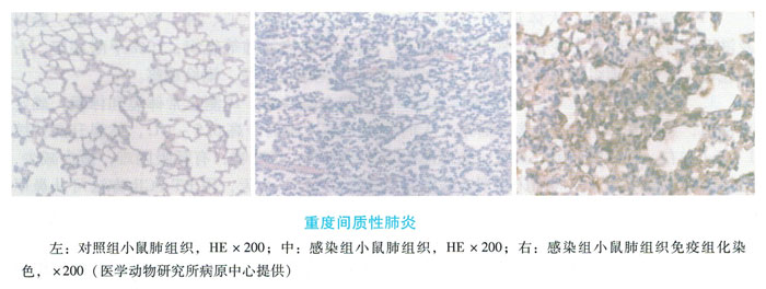
2. The morbidity and results of mice in the challenge group of BALB/c mice
infected by H5N1 model indicators: BALB/c mice began to decrease in activity on
the first day after challenge, refused to eat and shed hair, I was ready to die
from Starting on the third day, all died on the seventh day after the challenge.
T-PCR results show that there are multiple H5N1 viruses in lung tissues, and
virus isolation results show that the peak period of mouse virus replication in
mice is 2-6 days. After dissection, the mouse lungs can be observed to be
sclerotic, and the surface is dark red. The pathological changes showed focal
epithelial necrosis and shedding of the bronchial mucosa, obvious infiltration
of inflammatory cells in the mucosa, exudation in the alveolar cavity, and mild
expansion of the lung septum, resulting in severe interstitial. Cause pneumonia
(Figure 8-4).