Language

 动物造模,糖尿病大鼠视网膜病变
 z-bio
z-bio
 2021-10-07
 1054
1054
Background: Retinopathy remains one of the most devastating complications of diabetes. Basic science and clinical research have made great progress in understanding the complex pathophysiological mechanism of this blindness, but the exact mechanism is still unknown, and the effective treatment method is still unknown. Diabetic retina and capillary cells increase oxidative stress and may cause mitochondrial damage. In addition to capillary cells, other retinal cells, including photoreceptors and retinal pigment epithelial cells, have also been observed to increase oxidative stress. Antioxidants include lipoic acid, which supplements antioxidants and can prevent diabetic retinopathy in diabetic rats. In addition to oxidative stress, the retina also exhibits many abnormalities consistent with other inflammatory diseases. Vascular endothelial growth factor (VEGF) is an important angiogenic factor in vascular permeability and angiogenesis, and is elevated in the retina and vitreous of diabetic patients and animals. This increase is related to the symptoms of diabetic retinopathy. Redox-sensitive nuclear transcription factors-B and F-κB are activated, which is essential for the regulation of growth factor and cytokine expression, pro-inflammatory mediators such as interleukin 1β (IL-1β) and cell adhesion. Molecule 1 (ICAM-1) increases. Animal models indicate that antioxidants that prevent the development of diabetic retinopathy can prevent the increase in F-κB and IL-1β in the retina. Compared with other tissues, the high content of polyunsaturated fatty acids, coupled with the highest oxygen uptake and glucose oxidation, make the retina very sensitive to oxidative stress. The application of zeaxanthin to diabetic rats has been shown to prevent retinal oxidative stress and the increase of pro-inflammatory cytokines VEGF and ICAM-1. Antioxidants, including vitamin C and vitamin E, β-carotene, N-acetylcysteine and other micronutrients, can inhibit the development of diabetic retinopathy. The purpose of this study is to study the effect of nutritional supplements on diabetic retinopathy. An animal model of diabetic retinopathy was used to study the effects of multi-component nutrients on retinal capillary cell apoptosis, capillary degeneration and cell function. To study the effect of supplements on mitochondrial dysfunction, we quantified the gene expression of inflammatory cytokines, the DNA encoding cytochrome b and D1, and the expression levels of VEGF, IL-1β, and F-κB.
Method: Wistar rats (male, 200-225g) induced diabetes with streptozotocin and were divided into two groups. Many dietary supplements in rat group 1 (Prina 5001) contain carotenoids. eyepromiseDVS is specifically used to improve the structure and function of the retina, and is currently used in clinical trials of visual function in diabetic patients. Vitamin C nutrients (ascorbic acid, 300 mg), vitamin D3 (cholecalciferol, 10000 IU), vitamin E (vitamin E, 300 IU), fish oil ee 70 to 1.6 g), EPA (20 carbpentaenoic acid, 650 mg) , DHA (docosahexaenoic acid, 500 mg), benfotiamine (1 g), α-lipoic acid (750 mg), tocomin (200 mg), zeaxanthin (40 mg), lutein (20 mg), the patented formula contains 300 capsules of resveratrol. Green tea, turmeric, N-acetylcysteine, pine bark, grape seed extract, coenzyme Q10 zinc (2.65g), soybean oil. The second group of rats received Prina diet without supplements (DIAB) and used normal rats of the same age as the control group. During the entire study period (11 months), the average daily intake of diabetic rats was 50 grams. Eleven months after the start of the experiment, the rats were killed by carbon dioxide asphyxiation. One eye was suspended in 10 lm and digested with trypsin to prepare retinal capillaries, and the retina of the other eye was dissected to quantify conventional biochemical parameters. The liver was taken out and the absorption of some major components was confirmed by HPLC.
Retinal capillary cell apoptosis and histopathology: The retina was separated from formalin-fixed eyes and washed with water overnight. Digest the retina with trypsin containing 200 mM sodium fluoride for 45-70 minutes at 37°C to separate the capillaries. Apoptotic vascular cells were detected by terminal deoxyribonucleotide transferase preparation (TDT) using dUTP notch labeling staining (TUNEL method). Before the TUNEL reaction started, the retinal blood vessels were exposed to DNase as a positive control. Vascular and histological evaluation of TUNEL staining, Schiff periodate and hematoxylin staining. Count the number of acellular capillaries in the central area of the retina and express them as acellular capillaries in the retina.
Functional analysis: The retinal function of diabetic rats was measured 4 months later by measuring the electroretinogram (ERG). The rats were adapted to the night, anesthetized with ketamine and xylazine, and dilated their pupils with 1 pikaamide and 2.5 acid phenylephrine. Place the mouse on the heating platform and monitor the body temperature with a rectal thermometer. The A wave and b wave amplitude are measured by placing a silver eye on a threaded electrode embedded in a thin layer of cellulose passing through the surface of the cornea. The needle electrodes located on the tail and cheeks are used as ground electrodes and reference electrodes, respectively. Quantify reactive oxygen species (ROS) by fluorescence spectra of 2,7'-dichlorodiacetate. Incubate the protein (5-10 μg) with PBS containing 2uM DCHFDA for 10 minutes. Fluorescence is measured at wavelengths of 485m and 530m. Determine the antioxidant capacity of the retina. Mitochondrial Genome-Specific PCR Quantitative Elongation and 03XLPCR Kit determine the degree of mitochondrial DNA damage. The expression of VEGF, NF-κB and IL-1β was quantified by ELISA.
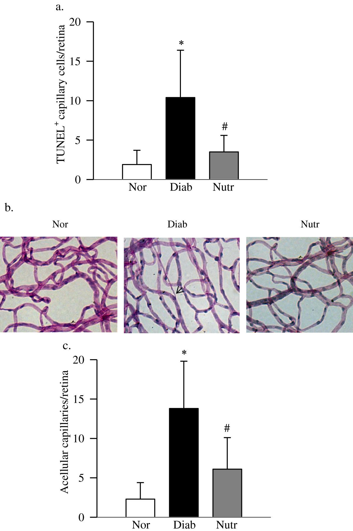
Results: Dietary supplements containing carotenoids prevent diabetic retinopathy from accelerating capillary cell apoptosis and histopathology: As expected, 4 out of 3 retinal vessels in 11-month-old diabetic rats TUNEL positive cells. Degenerate capillaries. Dietary supplements containing carotenoids can improve the apoptosis of capillary cells caused by diabetes. The number of TUNEL-positive capillary cells in normal diet rats is the same as that of diabetic rats. In diabetic rats treated with dietary supplements, the number of degenerative capillaries in retinal blood vessels was significantly reduced compared to rats without dietary supplements. Evaluation of retinal function by ERG measurement. Figure 2 shows that in diabetic rats with delayed electroretinogram response, the amplitude of A wave and B wave is significantly reduced. The decrease in the amplitude of the A wave and the B wave is due to the intake of a nutritionally supplemented diet.
Increased oxidative stress and mitochondrial damage in the retina of diabetic rats receiving dietary supplements: Compared with age-matched normal rats, the level of ROS in the retina of diabetic rats was significantly increased, and the total antioxidant capacity was also significantly reduced. However, compared with non-diabetic diabetic rats, supplemented diabetic rats had significantly lower OS levels and enhanced antioxidant capacity. Compared with diabetic rats not receiving dietary supplements, diabetic rats receiving dietary supplements can also prevent mitochondrial damage and significantly increase gene expression of mitochondrial DNA, which encodes a protein in the electron transport chain. Compared with normal rats, the levels of D1, ND6, and Cytb in the retina of diabetic rats were reduced by about 40-80? Diabetic rats receiving dietary supplements prevented this decline. use
Nutritional supplements can protect the retina from the increase of inflammatory mediators: the level of retinal vascular endothelial growth factor in diabetic rats is increased by 50? The increase in retinal vascular endothelial growth factor is improved by nutrients. Inflammation is considered to be the main cause of diabetic retinopathy.
Nutrition does not improve the severity of hyperglycemia in diabetic rats: Compared with unsupplied diabetic rats, the analysis of the main nutrients in the liver samples of nutrition rats is based on the time of α-tocopherol (36 μg/g to 74 μg/ g ), lutein increased by 3 times (0.02 μg to 0.06 μg), and zeaxanthin increased by 9 times (0.008 μg to 0.07 μg/g), indicating a 2-fold increase in levels.
Conclusion: Our data shows that for long-term diabetic patients, dietary supplements can protect nerve and blood vessel cells, inhibit the development of retinopathy, while maintaining the structure and function of the retina. I will. It can be achieved by improving the increase of inflammatory mediators and maintaining mitochondrial homeostasis, thereby protecting the retina from the self-diffusion virtuous circle of mitochondrial damage.