Language

 动物实验
动物实验
 z-bo
z-bo
 2020-08-12
 1029
1029
(1) Animal selection Choose healthy Japanese white rabbits or New Zealand
rabbits, either male or female.
(2) Surgical process According to different parts and methods of damaging
blood vessels, the required equipment and surgical procedures are different.
1. Surgical methods for different parts:
(1) Surgical preparation
1) Apparatus and medicine: ①Rabbit operating table (with rope for fixing).
②Surgery blade (big round knife), knife handle, round head surgical scissors,
ophthalmic scissors, ophthalmic forceps, dental forceps, vascular forceps, silk
thread, large angle needle, needle holder (the above surgical instruments need
to be sterilized). ③5% sodium pentobarbital, 1% lidocaine, 0.9% saline,
unfractionated heparin, 5% glucose solution.
2) Fixation and anesthesia: weigh the animal number and fix it on the
operating table. Slowly inject pentobarbital (0.5-0.7ml/kg) through the ear
vein, pay attention to the animal's breathing, and avoid respiratory depression.
The corneal reflex was tested, and the disappearance of the corneal reflex
indicated satisfactory anesthesia.
3) Skin preparation, disinfection, and drape: Cut off the hair of the
surgical field with curved shears, spread sterile towels and hole towels after
povidone iodine (iodine) disinfection, and fix them with towel forceps.
(2) Surgical procedure
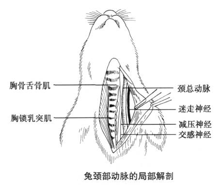
1) Common carotid artery (Figure 5-16): Fully expose the neck, touch the
common carotid artery to the right side of the trachea and determine the
position of the incision. Clamp the skin to both sides with dental forceps or
vascular forceps, use surgical scissors to cut a small cut on the skin between
the two vascular forceps, extend the vascular forceps into the incision, press
and lift the skin to open the vascular forceps to make it blunt Separate the
subcutaneous tissue, and then carefully cut the skin about 3 to 4 cm on the
upper and lower sides. Bluntly separate the subcutaneous tissue and neck muscles
and pull them outward to see the right common carotid artery sheath running
longitudinally on the right side of the trachea. In the sheath, the common
carotid artery accompanies the jugular vein, cervical vagus nerve, and
hypotensive nerve. The common carotid artery sheath can be separated first, and
then the common carotid artery can be separated from the sheath, and the
surrounding connective tissue can be removed to free the common carotid artery
with a length of 2 to 3 cm, as far as possible to the distal end. Follow-up
operations are performed according to different injury methods and surgical
purposes.
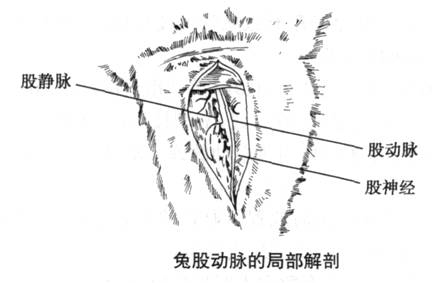
2) Femoral artery (Figure 5-17): Touch the strongest point of femoral artery pulsation and walk along the femoral artery. On both sides of the midpoint along the incision, use dental forceps or vascular forceps to clamp the skin to both sides, and use surgical scissors Cut a small cut in the skin between the two vascular forceps, extend the vascular forceps into the incision, press and lift the skin to open the vascular forceps to bluntly separate the subcutaneous tissue, and then carefully cut the skin about 2cm. The vascular forceps and fingers were bluntly separated to the femoral artery through the muscle space, and the femoral vein was seen running inside and below it. Select the unbranched blood vessel segment, carefully separate the loose connective tissue around the blood vessel to fully free the blood vessel. Follow-up operations are performed according to different injury methods and surgical purposes.
2. Injury method Air drying method and electric injury method are generally applied to the femoral artery and common carotid artery, and balloon strain and overdilation method are generally applied to the iliac artery.
The air drying method, electric injury method and balloon strain method (with or without high-fat feed) essentially make animal models of atherosclerosis. Various interventions are performed on the narrowed blood vessels within a certain period of time after surgery Treatment [balloon expansion and/or stent placement] to make a restenosis model (two-step method), of which balloon strain is the most widely used.
The rule of over-expansion is to create a restenosis model (one-step method) by damaging the arterial intima and media through over-expansion (balloon or stent) of normal blood vessels.
(1) Balloon strain method: The principle is to use a filled balloon to rub the surface of the vascular endothelium to cause endothelial exfoliation damage. With the aid of high-fat feed, it leads to the formation of lipid-rich plaques during the repair of arterial damage.
Equipment and equipment: Angiography machine, 0.014 inch guide wire, (2.0~2.5)×(15~20)mm balloon catheter, 5FJR angiography catheter, pressure pump, three-way three-way, Y-connector, Glucosamine meglumine
1) Atherosclerosis model: The surgical site is the femoral artery. Use the above method to fully free the femoral artery. Pass the double silk thread through the fully free femoral artery and ligate the distal end. The assistant pulls the proximal silk thread to block the arterial blood flow while pulling up the artery. The surgeon lifts the femoral artery with ophthalmic forceps, and uses ophthalmic scissors to make a 45° oblique incision on the artery wall 0.3cm on the proximal side of the distal ligature. The incision is about half the diameter of the tube.
Connect the balloon to the pressure pump and draw negative pressure. After inserting the tip of the 2.5mm×20mm balloon catheter into the arterial incision, the assistant loosens the pulled proximal silk thread and continues to insert the balloon catheter about 10cm, and expand the balloon at 3~5atm Pulling the balloon back after the capsule causes endothelial damage. After repeated 3 times, the catheter was removed and the artery was ligated. Stitched layer by layer.
2) Restenosis model: The surgical site is the common carotid artery. Use the above method to fully free the right common carotid artery. Pass the double silk thread through the fully free common carotid artery and ligate the distal end.
The assistant pulls the proximal silk thread to block the arterial blood flow while pulling up the artery. The surgeon lifts the femoral artery with ophthalmic forceps, and uses ophthalmic scissors to make a 45° oblique incision on the artery wall 0.3cm on the proximal side of the distal ligature. The incision is about half the diameter of the tube. After quickly inserting the 5FJR angiography catheter (the tip of the 0.014 inch guide wire is about 1mm from the tip of the catheter), the assistant loosens the pulled proximal end wire, continues to forward the guide wire and the catheter through the ligature, and then tightens the wire again.
Under the guidance of a 0.014 inch guide wire, the angiography catheter was sent to the abdominal aorta through the aortic arch and thoracic aorta, and the lower extremities arteriography was performed. 120U/kg of heparin was injected.
Withdraw the angiographic catheter, select the appropriate type of balloon and/or stent according to the reference vessel diameter and the length of the lesion, and expand the lesion with a stenosis of more than 50% at an appropriate pressure (usually 4 to 5 atm, 30 seconds/time × 3) , Record which side is expanding, the degree of lesion stenosis, lesion length, expansion pressure and the type of equipment used.
Repeat the radiography to ensure that the residual stenosis does not exceed 20%. If the stent is inserted, ensure that the stent adheres well.
Withdraw the guide wire and catheter, ligate the artery, and suture the wound layer by layer.
(2) Electrical stimulation method: The principle is that local electrical stimulation leads to vascular endothelial damage and may be accompanied by thrombosis, supplemented with high-fat diet to cause local atherosclerotic plaque formation.
Equipment and equipment: electrical stimulator, angiography machine, 0.014 inch guide wire, (2.0~2.5)mm×(15~20)mm balloon catheter, 5FJR angiography catheter, pressure pump, three-way three-way, Y-type connector, pan Meglumine
1) Atherosclerosis model: The surgical site is the common carotid artery or femoral artery. Use the aforementioned method to free the common carotid artery or femoral artery.
Place a stimulating electrode pad with autologous fat tissue on the proximal end of the blood vessel, and place the temperature electrode on the distal end. The distance between the two electrodes is about 1cm. The electrode hooks the blood vessel as gently as possible, and the surface is covered with wet saline gauze. Adjust the current to 1.2mA, stimulate for 12-15 minutes, and the degree of electrical damage should be local slight yellowing without obvious burn marks, local thinning, and proximal edema. Fix it next to the blood vessel with a metal clip as a marker. Suture the wound layer by layer.
2) Restenosis model: surgical site: if the surgical site of the atherosclerosis model is in the common carotid artery, the further angioplasty approach is in the ipsilateral external carotid artery, if the atherosclerosis model is in the femoral artery, the further approach is The angioplasty approach uses the right common carotid artery. The surgical procedure is the same as the balloon method.
(3) Air drying method: The principle is to use dry air to damage the vascular endothelium and cause thrombosis, supplemented with high-fat feed, locally forming atherosclerotic lesions.
Equipment and equipment: Angiography machine, 0.014 inch guide wire, (2.0~2.5)mm×(15~20)mm balloon catheter, 5FJR angiography catheter, pressure pump, three-way three-way, Y-connector, Glucosamine meglumine
1) Atherosclerosis model: The common carotid artery or femoral artery was freed using the aforementioned method. Pass the double silk thread under the fully free artery, the distance is about 1cm. Tighten the silk threads at the proximal and distal ends, and puncture the free segment of the artery with a 27G syringe needle. The puncture needle was used to dry and free the vascular cavity at a flow rate of 80 ml/min for a total of 10 minutes. After the saline is related to the free vessel segment, the ligature is removed. Fix it next to the blood vessel with a metal clip as a marker. Suture the surgical wound.
2) Restenosis model: surgical site selection: if the surgical site of the atherosclerosis model is in the common carotid artery, the further angioplasty approach is in the ipsilateral external carotid artery, and if the atherosclerosis model is in the femoral artery, further The angioplasty approach uses the right common carotid artery.
The surgical procedure is the same as the balloon method.
(4) Excessive expansion: The principle is that the local balloon and (or) the stent is over-expanded, which damages the vascular intima and media, and the local blood vessel overreacts to the injury to form restenosis.
Equipment and equipment: Angiography machine, 0.014 inch guide wire, (3.0~3.5)mm×(15~20)mm balloon catheter, 5FJR angiography catheter, pressure pump, three-way three-way, Y-connector, Glucosamine meglumine
Operation process: Use the above method to fully free the right common carotid artery.
Pass the double silk thread under the fully freed common carotid artery to ligate the distal end.
The assistant pulls the proximal silk thread to block the arterial blood flow while pulling up the artery. The surgeon lifts the femoral artery with ophthalmic forceps,
Use ophthalmological scissors to cut a 45° oblique incision on the artery wall 0.3cm on the proximal side of the distal ligation line. The incision is about half the diameter of the tube.
After quickly inserting the 5FJR angiography catheter (the tip of the 0.014 inch guide wire is about 1mm from the tip of the catheter), the assistant loosens the pulled proximal wire, and continues to send the guide wire and catheter forward, and pass the aortic arch and thoracic main under the guidance of the 0.014 inch guide wire The artery sends the angiography catheter to the abdominal aorta for arteriography of both lower extremities.
Inject 120U/kg of heparin.
Withdraw the angiographic catheter, select the balloon and/or stent according to the reference vessel diameter according to the ratio of 1.2 to 1.5:1, and send it to the femoral artery to expand and/or release the stent at an appropriate pressure (usually 8 to 14 atm), and record Which side is expanding, the pressure of expansion, and the type of equipment used.
Repeat the radiography and observe the final result. Withdraw the guide wire and catheter, ligate the artery, and suture the wound layer by layer.
(5) Precautions for surgery
1) Prepare 1% lidocaine, which can perform local infiltration anesthesia when the general anesthetic effect is not satisfactory, reduce the animal's struggle, and also avoid excessive intravenous anesthetic resulting in animal death; in addition, after fully freeing the blood vessel segment, Before the intubation operation, lidocaine can be infiltrated locally, and the blocking of the local vascular motor nerve can make the blood vessel dilate, which is convenient for the intubation operation.
2) Rabbits have poor tolerance to blood loss, and should be minimized by skilled surgical operations. If blood loss is high, normal saline or 5% glucose solution can be used to supplement blood volume.
3) It is difficult for the catheter to pass through the aortic arch to the thoracic aorta, and attention should be paid to the shaping of the tip of the guide wire.
4) Two-step animal model, 4 weeks after making the atherosclerosis model, the restenosis model is made.
(3) Postoperative animal handling and specimen selection and fixation
1. Postoperative treatment: Postoperative intramuscular injection of 800,000 U/d of penicillin for 3 consecutive days. High-fat feed contains 2% cholesterol and 6% peanut oil. The over-expansion animal model is fed only ordinary feed.
2. Fixing and taking materials
(1) Fixation and material extraction are performed four weeks after operation.
(2) Equipment and equipment: angiography machine, 0.014 inch guide wire, 5FJR angiography catheter, diatrizoate meglumine; rabbit operating table (fixed rope); surgical blade (large round knife), knife handle, round head surgical scissors, ophthalmology Scissors, flat forceps, vascular forceps, silk thread, 7F arterial sheath, infusion set, specimen bottle; 5% sodium pentobarbital, 0.9% saline, 4% formaldehyde.
(3) Operation process (take the iliac artery restenosis model as an example): fix the animal by anesthetizing as described above, separate the left common carotid artery and intubate it to the bifurcation of the abdominal aorta, perform bilateral iliac angiography, record the images,备Measurement.
Open the abdomen layer by layer in the middle to the abdominal cavity, push the large colon to one side, and bluntly separate the mesenteric and posterior peritoneum from the loose connective tissue to the abdominal aorta. Separate the abdominal aorta in front of the spine and the abdominal vena cava on the left side of the spine clearly and free them separately, and then pass a double silk thread under it, and ligate both proximal to the heart.
The assistant pulls the distal silk thread to block the blood flow while pulling up the artery. The surgeon lifts the blood vessel with flat forceps, and makes a 45° oblique incision on the vessel wall with ophthalmic scissors. The incision is about half the diameter of the tube. Insert one arterial sheath into each, and ligate the distal end of the silk thread to fix the sheath.
Move the rabbit operating table to the sink, connect the side tube of the abdominal aorta sheath with normal saline, open the side tube of the abdominal venous sheath, the liquid level of the perfusion bottle (bag) is 140-150cm higher than the animal, and the flow rate of the perfusion solution is 2.0-2.5 ml/min. When the venous effluent becomes clear, 4% formaldehyde is used for in situ physiological pressure perfusion, and each rabbit is perfused with 100-200ml perfusion fluid.
Separate and intercept the target blood vessel segment and place it in a 4% formaldehyde sample bottle for fixation.
