Language

 心肌梗死模型
 z-bo
z-bo
 2020-08-12
 1050
1050
1. Selection of Experimental Animals In previous studies, models of myocardial infarction have been established in various experimental animals. When small animals can be selected according to the requirements of the research, mice are the best choice. On the one hand, the cost of buying and raising mice is relatively low, and on the other hand, because molecular biology tools (such as gene knockout and genetically modified animals, etc.) are more used for mice , Especially mice. Dogs, sheep, and pigs have all been used when large animals need to be used as experimental subjects according to the requirements of the research, but for the reasons detailed in the previous section, pigs are currently the most ideal choice. When an animal model of acute myocardial infarction is made by ligation and other methods, the animals with abundant collateral circulation such as dogs are generally not suitable for use. In these animals, due to the abundant original collateral vessels, the coronary arteries are acute After occlusion, the collateral circulation opens rapidly, and large-scale myocardial infarction is rarely formed, which is difficult to meet the requirements of the research.
2. Model building method
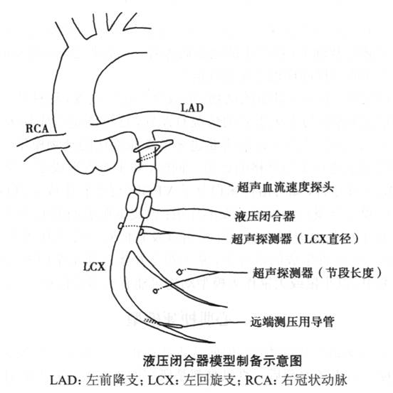
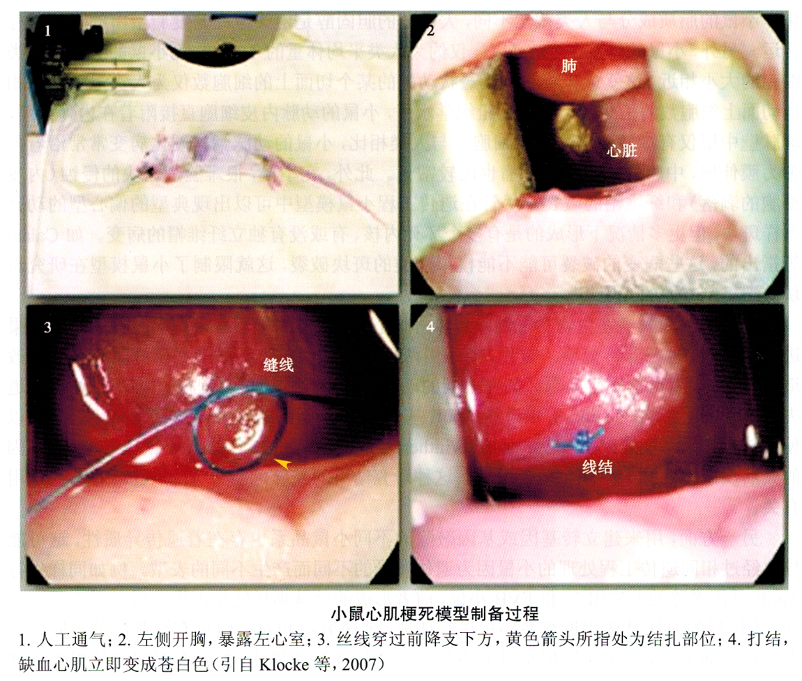
(1) Coronary artery branch ligation (Figure 6-6, Figure 6-7): The most direct method for acute coronary occlusion is to open the chest to ligate the coronary artery branches. This model was first proposed by Selye et al. in 1960. It can be applied from childhood Various experimental animals such as rats, rats, rabbits and pigs. This model has been repeatedly verified by many researchers from the aspects of histology, anatomy and function, and has been used in pathophysiological research and drug evaluation. At present, this model is mainly used to study the long-term results of acute myocardial infarction, such as ventricular remodeling and heart failure. Due to the differences in the anatomy of the coronary arteries between individual animals and the differences in the selection of ligation sites during surgery, the level of coronary artery branch ligation is difficult It is completely consistent. When making the model, the range of myocardial infarction will have a large variation, which can account for 15% to 60% of the ventricular muscle. In order to obtain sufficient statistical significance, a larger sample size is required. Therefore, when allowed Try to choose small animals for research.
(2) Coil embolization: In large animal models, intravascular coils can be used to cause acute occlusion of coronary artery branches. Through carotid or femoral artery puncture, the coil is placed in the distal segment of the three main coronary arteries or the starting segment of the large branch by intravascular intervention. The advantage of using this method is that thoracotomy is avoided, and the model can be prepared by intravascular intervention. However, compared with ligation under direct vision, the vascular occlusion is not accurate enough and the range of intravascular thrombosis is not easy to control. Such coils are commercially available, and are often used for embolization of peripheral arterial hemangioma, but the cost of using commercially available products is high. In animal models of acute myocardial infarction, the application of this model is less.
(3) Coronary artery microembolism: There have also been researchers who used drugs (such as isoproterenol) or microembolization to create diffuse micro myocardial infarction. For example, Medvedev used plastic microspheres with a diameter of 159m to be directly injected into the coronary artery. Embolization of the vascular bed within a certain range, causing experimental heart failure.
In fact, acute myocardial infarction models prepared by surgical methods are more used in the study of heart failure after myocardial infarction, and it is difficult to provide useful information for research on the prevention and treatment of human myocardial infarction. Therefore, it is more important to establish an animal model of spontaneous myocardial infarction that is similar to the pathophysiological process of human myocardial infarction.
