Language

 动物实验,H5N1病毒
 z-bo
z-bo
 2020-08-12
 1079
1079
1. Serological screening of ferrets Ferrets undergo serological screening for influenza before infection, and at least the animals whose serological test results for influenza virus are negative can be included in the group.
2. Materials and methods
(1) Experimental virus: Allantoic fluid harvested from SPF chicken embryo passage. The TCID50 of the virus liquid was determined to be 10 to the 8th power/ml.
(2) Animals and infection methods: Ferrets aged 4-10 months, weighing 500-1000g, serum antibody test should be carried out before infection. Ferrets that are negative for antibodies to H5N1 virus and influenza that are immediately circulating are qualified and can be selected for experiment . Ferrets were inoculated with 100 TCID50 (500 μl volume) through the nose. All animal experiments were performed in the ABSL-3 laboratory. On the 5th day of infection, the ferrets were euthanized and collected.
(3) Clinical observation: Before the experiment of ferrets, a chip was implanted under the skin, the body temperature was read through the chip once a day, and the body weight was weighed once a day to observe for cough, runny nose, vomiting, diarrhea, shortness of breath, dyspnea, and obvious loss of appetite And other clinical manifestations, detailed records and records.
(4) Virus isolation: 1 day after virus inoculation, turbinate bone biopsy specimens are collected every day and placed in 1ml DMEM culture medium for use. The specimen was inoculated with MDCK cells and adsorbed for 1 hour for virus isolation. Ferrets were euthanized to death on the 5th day after infection. Lung, trachea, liver, spleen, brain, intestine, heart and kidney tissues were taken during autopsy, and after grinding treatment, MDCK cells were inoculated. Observe cytopathic changes every day for 3 days, and confirm HA by turkey red blood cells. The HA method is specifically as follows: Take out 50 μl of the MDCK cell supernatant of the inoculated specimen, add an equal volume of turkey red blood cells, and react at room temperature for 30 minutes. Agglutination occurs, and the result is positive.
(5) Detection of H5N1 virus RNA: 1 day after H5N1 virus inoculation of animals, turbinate bone biopsies are collected every day, and viral RNA is extracted in the ABSL-3 laboratory. Operation steps: 100 μl of turbinate bone biopsy specimens are transferred into the lysis solution (Qiagen ), put 350μl of buffer RLT (with 1% mercaptoethanol added), centrifuge at 13000rpm at 4°C for 3min, transfer the supernatant to a new 1.5ml microcentrifuge tube, and extract viral nucleic acid from the turbinate bone biopsy sample. The RNA was reversed into cDNA, and the viral load was directly measured by a fluorescent quantitative PCR machine.
(6) H5N1 virus antibody detection: After the animal is sacrificed, blood is collected to separate the serum and stored at -80°C for later use. Serum H5N1 viral antibody detection (H1 method) The specific operation steps are: add 1:10 to 1:1280 diluted serum to be tested to each well, add 8HA virus, and incubate at room temperature for 30 min. Afterwards, the result of agglutination is tested, and the titer of serum agglutination is the titer of antibodies that can be neutralized by the serum.
(7) Pathological research: general observation, after the animals were sacrificed, the morphological changes of lung, heart, spleen, kidney, liver, brain, intestine, and lymph nodes were observed in detail; pathological section preparation, and lungs and other tissues of the sacrificed animals were taken. Fixed in% formaldehyde, embedded in paraffin, treated with conventional pathological tissue sections, stained with HE or immunohistochemistry, and observed under microscope.
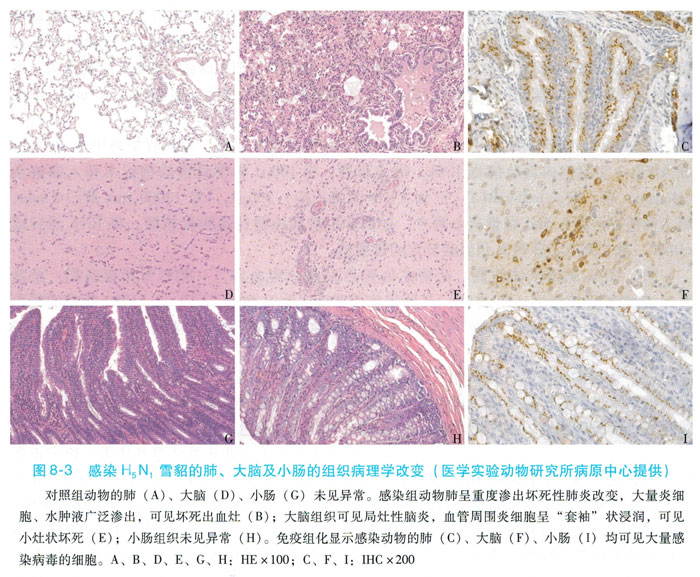
3. Ferrets infected with H5N1 model indicators and results. Ferrets were tested with normal body temperature and body weight one week before the experiment to determine the normal values. The temperature of ferrets increased on the second day of H5N1 virus inoculation, and the body temperature increased by 0.5-2℃. The increase lasted for about 3 days, and there was clinical death. After being infected, ferrets have clinical manifestations such as runny nose, sneezing, difficulty breathing, reduced food intake, lethargy, decreased activity, and no response to external stimuli. From day 1 to day 5 after the virus infects ferrets, the viral load can be detected from the turbinate bone biopsy by RT-PCR. From the turbinate bone specimens and lung tissue specimens of ferrets, the MD-CK cells were cultured and the cells were inoculated to produce typical cytopathic changes (CPE), suggesting that the ferret was infected with the virus and the virus was replicated and detoxified. The turbinate bone specimens were isolated positive The peak period is 2 to 6 days. Live viruses can be isolated from brain, heart, liver, spleen, intestine, kidney, and lymph node tissues. This result proves that H5N1 virus can replicate and reproduce in tissues and organs other than the respiratory tract. The HA method can confirm the presence of H5N1 antigen in MDCK cells. Before H5N1 virus inoculation and on the 5th day after inoculation, 1ml of blood was collected from the vein to separate the serum, and the H5N1 virus-specific antibody in the serum was detected. The infected ferrets were all negative when they were euthanized for 5 days. General anatomical observations can reveal mild consolidation on the surface of the lung tissue, and microscopic examination shows obvious inflammatory exudates in the alveolar cavity, accompanied by lymphocytes and phagocyte infiltration. Extensively widened pulmonary septum with infiltration of a large number of inflammatory cells dominated by monocytes, alveolar deformation becomes smaller under compression, and there are limitations in alveolar septum widening and then fused with each other, and large flaky alveolar septa widened and fused with each other in some areas. There are lymphocytes, plasma cells, and phagocytes in the alveolar compartment, with a wide range of lesions, showing moderate to severe interstitial pneumonia, as shown in Figure 8-3.