Language


 bd-xu
bd-xu
 2021-12-21
 976
976
As we all know, the new coronavirus (CoV) is named as COVID-19 virus, which has been rampant in the world for nearly half a year, seriously harming human life and health and hindering the economic development of society. However, there is still no targeted treatment. Many studies on the epidemiology and clinical characteristics of COVID-19 have been published. However, due to its human-to-human infectivity and the mutation of genetic material, it is almost impossible to perform autopsy or biopsy in the early stage. The biopsy makes the pathological information of this disease very scarce, which makes it impossible to provide new ideas for clinical treatment. In order to fill the vacancy in this regard, in recent months, Chinese scholars have published some pathological research reports of patients with new coronary poisons after being authorized.
Histopathological results of 4 middle-aged and elderly patients with COVID-19 pneumonia in Zhongnan Hospital of Wuhan University
On April 14, Dr. Liao Meiyan from the Department of Radiology, Central South Hospital of Wuhan University, and Dr. Xiao Shuyuan, a pathologist at the University of Chicago, and his team published the topic Pathological study of the 2019 novel coronavirus disease (COVID-19) through postmortem core biopsies on Modern Pathology The medical research report published the histopathological results of the autopsy of 4 patients died of new coronavirus, which provides new ideas for the development of treatment plans.
The experiment performed post-mortem lung, liver and heart biopsy on 4 patients with COVID-19 pneumonia aged between 59 and 81 years. Each patient has at least one underlying disease, including an immunodeficiency state (chronic lymphocytic leukemia and kidney transplantation) or other conditions (cirrhosis, hypertension, and diabetes), and the time from onset to death ranges from 15 days to 52 days. Examination found that all patients' white blood cell counts increased, and increased significantly towards the end. Except for patients with leukemia, all patients showed lymphopenia. Histopathological observations were mainly manifested in the lungs, including alveolar epithelial cell damage, clear membrane Formation and type II lung cell hyperplasia are all components of diffuse alveolar injury. Fibroblast proliferation, extracellular matrix and fibrin accumulation in the air are obvious.
The study found that the pathological basis of COVID-19 pneumonia is some patients with advanced DAD (organization/fibrosis) and over-infected bacterial pneumonia. Early organization is obvious but there is no obvious fibrosis. In conclusion, necropsy revealed late diffuse alveolar injury, some patients with bacterial pneumonia, liver and heart changes may be secondary, or related to underlying disease.
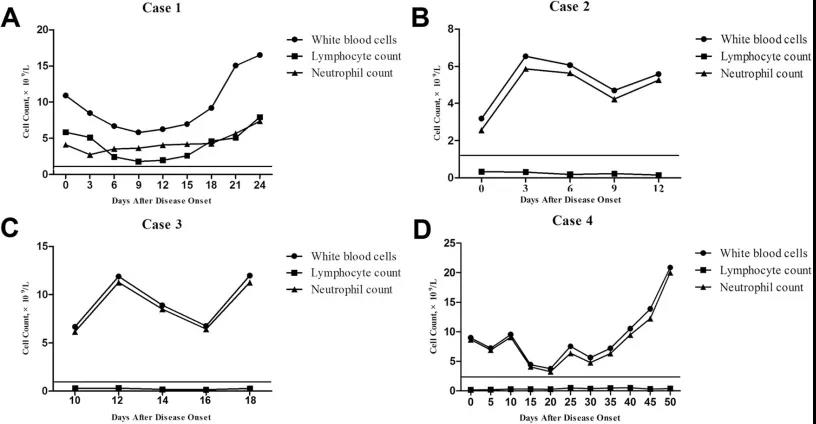
Count of white blood cells, neutrophils and lymphocytes in 4 patients in time
(A) Case 1 increased lymphocyte count consistent with her history of chronic lymphocytic leukemia;
(B, C, D) Cases 2, 3 and 4 have markedly reduced lymphocytes
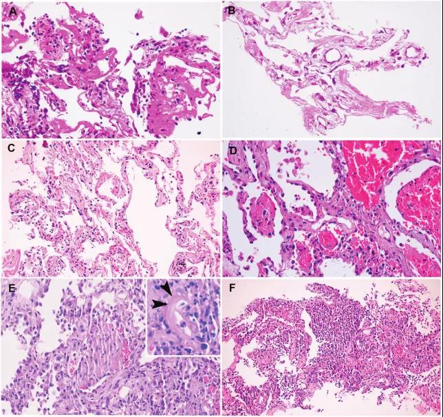
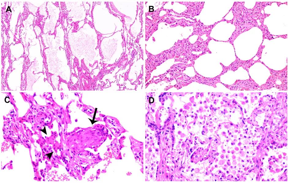
Lung histopathological observation
(A) Case 1: Thick glass-like membrane with desquamative lung cells and mononuclear inflammatory cells;
(B) Case 2: The hyaluronic membrane is delicate and there is no obvious inflammatory infiltration;
(C) Case 3: Focal transparent membrane, type II lung cell hyperplasia, mild interstitial thickening;
(D) Case 4: The alveolar cavity is filled with red blood cell exudate, and small fibrin plugs can be seen in the adjacent alveoli;
(E) Organization of fibroblasts mixed with fibrin and inflammatory cell infiltration in alveoli, diffuse lung cell hyperplasia in the background (inset: fibrinoid vascular necrosis, indicated by arrows;
(F) Bronchopneumonia is accompanied by marked changes in neutrophil infiltration, and the alveolar cavity is filled.
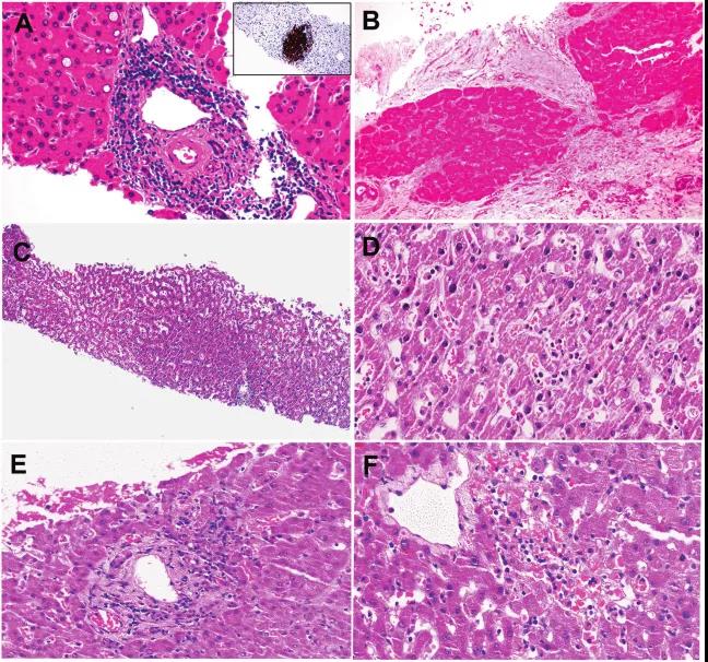
The liver pathology of 4 patients
(A) Case 1 Atypical small lymphocytes (insertion: CD20 immunostaining) with dense portal infiltration and focal glycosylated nuclei in hepatocytes;
(B) Case 2 cirrhotic nodules with thick fibrosis;
(C) Mild sinusoid expansion with increased lymphocyte infiltration;
(D) High power microscope shows lymphocytes;
(E) Focal liver necrosis around the portal vein;
(F) Case 4 had focal central hepatic lobular necrosis.
Pulmonary adenocarcinoma patients with early COVID-19 lung disease histopathological report
On February 20th, Dr. Xiaoshu Yuan and his team published a paper entitled Pulmonary pathology of early-phase 2019 novel coronavirus (COVID-19) pneumonia in two patients with lung cancer in the Journal of Thoracic Oncology, reporting 2 cases Pulmonary adenocarcinoma patients with early COVID-19 lung pathology changes. Pathological examination revealed that in addition to the tumor, both patients had pulmonary edema, protein-like exudate, focal reactive lung cell hyperplasia with inflammatory cell infiltration, patchy, and multinucleated giant cell infiltration. The transparent film is not obvious. Neither patient showed signs of pneumonia at the time of surgery, and these changes may represent the early stages of pulmonary pathology of COVID-19 pneumonia.
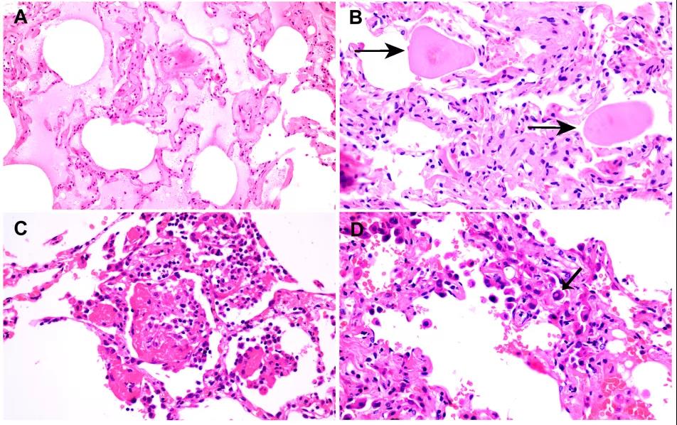
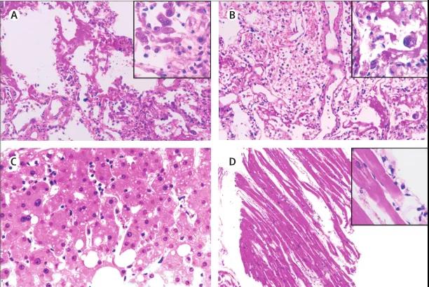
Case 1 histopathological changes of COVID-19 pneumonia
(A) Exudate in the alveoli, with particles;
(B) scattered large protein balls (arrows);
(C) Early fibrin tissue in the alveoli, with mononuclear cells and multinucleated giant cells;
(D) Hyperplastic lung cells with suspicious viral inclusions (arrows).
Case 2 Histopathological changes of COVID-19 pneumonia
(A) Obvious protein and fibrin exudates;
(B) Fibroblast hyperplasia and type II lung cell hyperplasia lead to diffuse expansion of the alveolar wall and septum, consistent with the early pattern of diffuse alveolar injury (DAD);
(C) Embolization of fibroblasts or fibroblast balls proliferating in the stroma (arrow);
(D) A large number of macrophage infiltration and type II lung cell proliferation.
Pathological results of the first COVID-19 patient's body
On February 17, Academician Wang Fusheng and Professor Zhao Jingmin of the Fifth Medical Center of the Chinese People's Liberation Army General Hospital and their team published a report entitled "Pathological findings of COVID-19 associated with acute respiratory distress syndrome" on Lancet Respir Med. The results of the first pathological examination of the remains of COVID-19 patients mainly introduce the diagnosis and treatment of a new crown patient and the pathological manifestations of COVID-19-related acute respiratory distress syndrome: the lung pathology of the patient at the time of death suggests that he is in the exudative period of DAD No obvious organizing and pulmonary fibrosis were found in lung pathology.
Pathological manifestations of right (A), left (B) lung tissue, liver tissue (C), and heart tissue (D) in patients with severe pneumonia caused by SARS-CoV-2
Overall, the pathological analysis of the new crown patients reported above can not only provide new insights into the pathogenesis of SARS-CoV-2 related pneumonia, but also help doctors formulate timely treatment strategies for such severe patients and reduce mortality. At the same time, in order to further understand the pathogenesis of COVID-19, we need more patients of different ages and different physiological backgrounds to participate in the study. In addition, it is necessary to establish appropriate animal models, not only to imitate the infection itself, but also to imitate the pattern of human disease progression, so as to better and faster overcome the virus.
▎References:
https://doi.org/10.1016/S2213-2600(20)30076-X
https://doi.org/10.1016/j.jtho.2020.02.010
https://doi.org/10.1038/s41379-020-0536-x
